Structure of ZIKV
Similar to other flaviviruses, ZIKV is an enveloped, icosahedral virus with a non-segmented, single-stranded, positive-sense RNA genome, closely related to the Spondweni virus. The genome is 10,794 bases long, and has two non-coding flanking regions, the 5′ NCR and the 3′ NCR. It comprises a single long open reading frame: 5′-C-prM-E-NS1-NS2A-NS2B-NS3-NS4A-NS4B-NS5-3′. The NS1, NS3, and NS5 are large, highly-conserved proteins, while the NS2A, NS2B, NS4A, and NS4B proteins are smaller, hydrophobic proteins. The ORF codes for a polyprotein that subsequently cleaves into the capsid (C), precursor membrane (prM), envelope (E), and non-structural proteins (NS) [11].
The 5’ end has a methylated nucleotide cap. The non-polyadenylated 3’ terminus has a looped structure. The secondary structure leads to the formation of a sub-genomic flavivirus RNA (sfRNA), which is essential for pathogenicity [12].

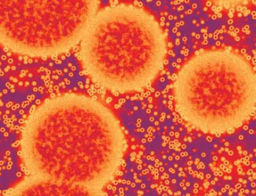
Fig 3: Virion. Enveloped, spherical, about 50 nm in diameter. The surface proteins are arranged in an icosahedral-like symmetry. Source: Zhang et al
The E protein composes the majority of the virion surface and is involved with aspects of replication, such as host cell binding and membrane fusion. Cryo-electron microscopic studies showed ZIKV to be similar to other flaviviruses. The envelope (E) protein is very similar to the West Nile virus, Japanese encephalitis virus, and the dengue virus [13].
The virion is about 40 nm in diameter, having 5-10 nm surface projections arranged in an icosahedral symmetry. The ZIKV contains a nucleocapsid (approx. 25-30 nm in diameter), surrounded by a lipid bilayer, containing the prM and E envelope proteins [13].

Fig 4: Genome Source: http://viralzone.expasy.org/all_by_protein/6756.html
Replication of ZIKV
Using the PF-13 ZIKV strain isolated during an outbreak in French Polynesia, Hamel et al. extensively studied the interaction of ZIKV in humans[14]. They observed that fibroblast, keratinocytes, and immature dendritic cells are all susceptible to ZIKV infection, and support viral replication. Once the virus is deposited on the epidermis and dermis of the host, there is a rapid increase in RNA copy numbers and ZIKV particles, which indicate active viral replication. The ZIKV infection of the epidermal keratinocytes results in cell apoptosis, indicated by the appearance of cytoplasmic vacuolation and the presence of pyknotic nuclei in the stratum granulosum. Inducing apoptotic cell death prevents the antiviral immune response, and increases the dissemination of the virions. The E protein is responsible for the fusion and entry of the virus through the host membrane. However, the exact mode of entry is not yet understood.
Post dissemination, the virus continues to spread to the lymph nodes and blood stream. Although flaviviruses are generally known to replicate in the cytoplasm, ZIKA antigens have also been detected in the nuclei of infected cells [15].


